Chapter 17 – Ophthalmic System Assessment – The Eyes
Peripheral Vision Assessment
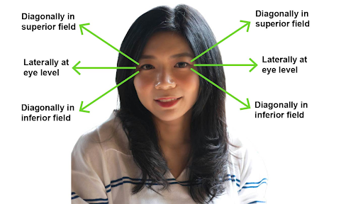
Peripheral vision can be assessed using the confrontational visual field exam. This exam can be conducted in several ways including client peripheral vision compared to examiner, the wiggle finger method, and the counting finger method. For each of these tests, move your arm/hand into three of the client’s peripheral vision fields as shown in Figure 20: superior, eye level, and inferior.

Figure 20: Peripheral vision fields.
Conduct these assessments in well-lit environments with the client’s eyes at the same level as yours. Both of you may stand if your eye levels are similar. Alternatively, you can stand and then raise or lower the exam table so that the client’s eyes are at the same level as yours.
Ask the client to remove anything that could interfere with peripheral vision such as a hat with a rim. Additionally, if they have a loose head covering, they may need to tighten/tuck it behind their ears. Ask them to look at your face and whether they can clearly see you with no difficulty and no blurring.
Then, follow these steps for each of the assessments:
- Client peripheral vision compared to examiner: Stand about one foot away from the client. Cover your own right eye and ask the client to cover their left eye with the palm of their hand and stare directly at your open eye; you will also stare at their open eye. Stretch your left arm/hand out laterally as far as you can and diagonally into the superior field, at the client’s eye level, midway between you and the client. Begin to move your arm/hand inward. Ask the client to state “now” when they first see your hand: they should see it at the same time you see it. Repeat this by stretching your arm/hand diagonally into the inferior field and moving your arm/hand inward. Then, repeat a third time with your arm/hand stretched out laterally at the client’s eye level, but this time closer to them as opposed to midway between you and the client. Then, repeat the same three steps on the opposite eye.
-
- Normally, the client’s peripheral vision should equal the examiner’s vision. Be aware that the validity of this exam is based on the assumption that you as the examiner have normal peripheral vision, which is about 90 degrees at the superior and inferior field, and a bit greater than this at the eye level.
- Abnormal findings are when the client has decreased peripheral vision such as less than 90 degrees.
For the Wiggle finger method and the counting finger method, position yourself about 3–4 feet away from the client.
- Wiggle finger method: Ask the client to fixate (focus) on your nose. Stretch both of your arms out to the side, pointed diagonally into the superior quadrants. Wiggle/flex the index finger on one of your hands and ask the client to point to the side that they see your finger moving. Then repeat with your other hand. Repeat the same steps with your arms stretched out to the side laterally at eye level and then out to the side pointed diagonally into the inferior quadrants (see Video 6).
- Normally, the client should be able to identify the correct hand that is moving each time.
- Abnormal findings are when the client reports not seeing the hand wiggle and/or not being able to identify the correct hand that is moving each time.
Video 6: Wiggle finger method. [0.45 seconds].
- Counting finger method: To test the client’s right eye, cover your right eye and ask the client to cover their left eye with the palm of their hand and stare directly at your open eye; you should do the same. Now, stretch your left arm/hand out pointed diagonally into the superior quadrant, then laterally out at the eye level, and then diagonally down into the inferior quadrant. In each of these fields, hold up 1–5 fingers (use a different number each time) and ask the client how many fingers they see. Your hand should be midway (equidistant) between you and the client (i.e., not closer to you, nor closer to them) with the palm of your hand facing the client, so they can tell how many fingers you are holding up. Repeat the same steps to test the client’s left eye; this time asking them to cover their right eye while you cover your left and use your right arm/hand. See Video 7 for an example of testing the right eye.
- Normally, the client should be able to accurately report how many fingers are held up in the superior, lateral/eye, and inferior field on both sides.
- Abnormal findings are when the client has difficulty seeing the fingers and/or has difficulty or is unable to identify how many fingers are being held up.
Video 7: Counting finger method for testing the right eye. [0.42 seconds].
- Note the findings:
- Normal findings might be documented as: “Confrontational visual field exam: client able to identify correct numbers and hand wiggling in all three peripheral positions on left and right sides. Client’s peripheral vision intact (equal to the examiner’s).”
- Abnormal findings might be documented as: “Confrontational visual field exam: client unable to see correct numbers displayed by examiner’s fingers and examiner’s hands wiggling in superior and inferior fields bilaterally. Client’s peripheral vision is less than the examiner’s.”
Priorities of Care
Clients with decreased peripheral vision should be encouraged to see an eye specialist.
However, new onset and sudden loss of peripheral vision such as a dark spot or shadow requires emergency care as these symptoms could be related to a stroke or . In these cases, notify the physician or nurse practitioner immediately, stay with the client, take their blood pressure, and perform a primary survey.
is an emergency situation when the retinal tissue pulls away from the back of the eye (without immediate treatment it can result in permanent loss of vision).

