
Chapter 15 – Neurological system assessment
CN V – Trigeminal Nerves
- Sensory function testing of CN V – Cotton ball: Explain to the client that you want them to close their eyes, that you will lightly touch areas on their face with a cotton-tipped applicator or cotton ball, and that they should indicate when they feel you touch their face each time. When their eyes are closed, briefly touch one spot in the ophthalmic area, then wait 2 to 3 seconds, and then touch the other ophthalmic area (see Figure 13). After touching these two spots, ask the client if the sensation felt the same. Repeat the same test for the maxillary and mandibular areas. The objective is to evaluate the locations where the trigeminal nerves innervate (see Figure 14 and Video 7).
-
- Normally, a client should be able to indicate when their face is being touched in the ophthalmic, maxillary, and mandibular areas bilaterally and the sensation should feel similar.
- Abnormal findings include not being able to feel the touch on one or more locations or having decreased sensation on one side. If you observe any abnormal findings or need more information, you may repeat the test using a tongue depressor broken in half. Apply the rounded edge or sharp edge in each location and ask the client to differentiate between “rounded edge” or “sharp edge.” You may show them the round edge and sharp edge before beginning and brush both on their forearm so they know what each feels like.
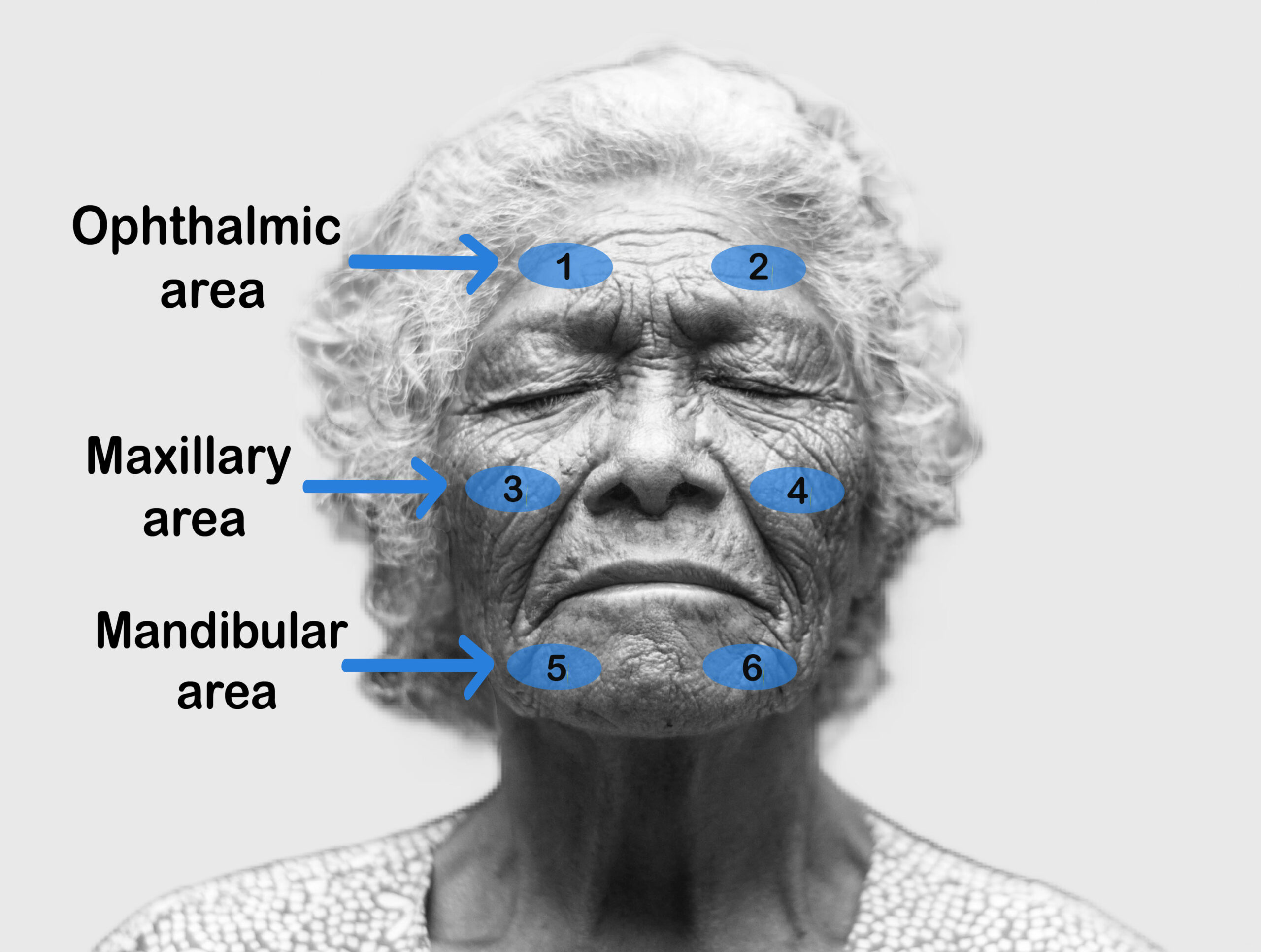
Figure 13: Trigeminal nerve sensation areas. (Illustrated by Tayiba Rahman)
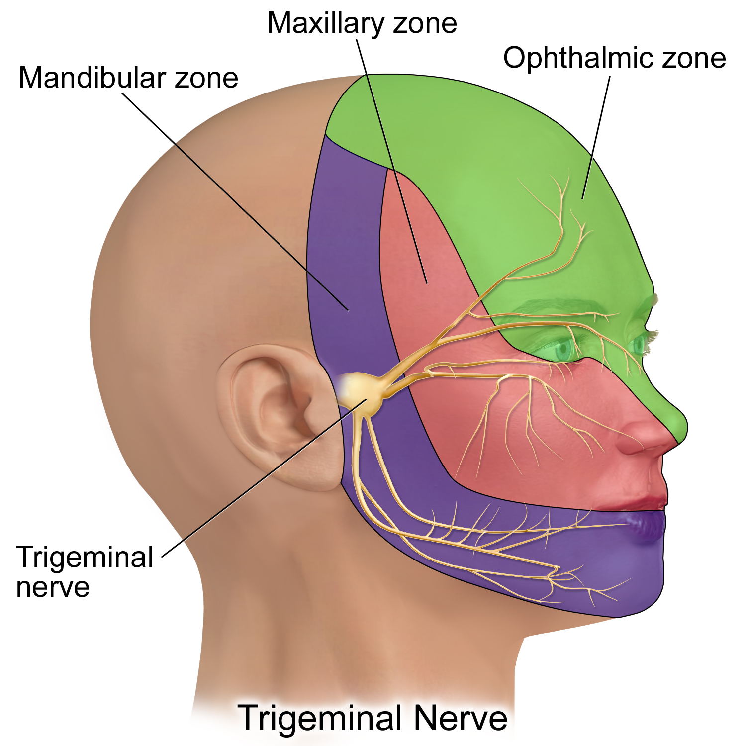
Figure 14: Sites of trigeminal nerve innervation. (CC-BY 4.0 by BruceBlaus: https://commons.wikimedia.org/w/index.php?curid=61465350)
Video 7: Sensory testing of trigeminal nerve (CN V).
- Sensory function testing of CN V – Corneal reflex: Test the corneal reflex ONLY in specific situations, such as suspected brain stem damage or CN V or CN VII lesions – typically, the corneal reflex should NOT be tested on a person who is healthy. The corneal reflex is facilitated by afferent sensory CN V and efferent motor CN VII. To test the right eye, stand on the client’s right side; if they are able to follow commands, ask them to keep their head still to use their eyes only to look up and away from you. Then, with a delicate wisp of a cotton ball, gently touch the edge of the cornea. Repeat this procedure for the left eye. Some healthcare professionals perform the test with a drop of sterile saline instead of a wisp of the cotton ball to avoid possible corneal abrasion. See Video 8.
- Normally, there will be bilateral blinking.
- Abnormal findings include unilateral or absent blinking.
Video 8: Corneal reflex.
- Motor function testing of CN V – Inspect and palpate: Inspect the temporalis and masseter muscles for symmetry. Next, ask the client to clench their teeth while you palpate the temporalis and masseter muscles on both sides simultaneously along the upper and lower jaw.
- Normally, the muscles are equal bilaterally in size.
- Abnormal findings include asymmetrical muscle mass or atrophy.
- Motor function testing of CN V – Range of motion: Ask the client to open and close their mouth a bit slower than usual and then protrude and retract the lower jaw (move outwards and back inwards).
- Normally, these movements should all be symmetrical.
- Abnormal findings include asymmetrical movement.
- Motor function testing of CN V – Manual muscle testing: Perform manual muscle testing of the jaw. Ask the client to keep their head still and clench their teeth, and then place your thumbs on their chin with your fingers toward the back of the neck. With your thumbs, attempt to pull down their lower jaw. Then, ask them to open their mouth and resist your force (i.e., not let you push their jaw closed) while you apply upward pressure on their chin; to do so, place your fingers on their chin applying upward pressure and your other hand on the top of their head (see Video 9).
- Normally, the client should be able to resist your force, demonstrating good strength.
- Abnormal findings include decreased or no resistance in which the client cannot resist jaw opening unilateral or bilateral, or is unable to keep the jaw open, or the jaw deviates to one side.
Video 9: Manual muscle testing of the jaw.
- Note the findings:
- Normal findings might be documented as: “Trigeminal nerve test: sensations intact on ophthalmic, maxillary, and mandibular areas bilaterally. Temporal and masseter muscles equal in size, symmetrical movement observed with ROM. Client able to resist force on the jaw. Corneal reflex present bilaterally.”
- Abnormal findings might be documented as: “Trigeminal nerve test: no sensations felt on ophthalmic, maxillary, and mandibular areas bilaterally, temporal and masseter muscles atrophy bilaterally, no muscle strength in jaw, corneal reflex absent bilaterally.”
