Chapter 17 – Ophthalmic System Assessment – The Eyes
Extraocular Eye Movement
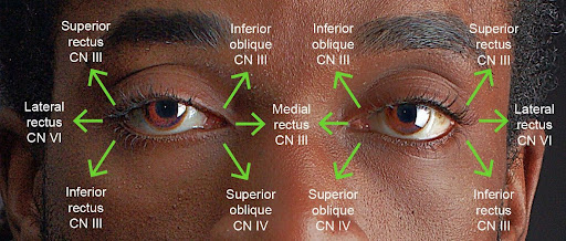
The extraocular eye movement is innervated by:
- CN III (oculomotor).
- IV (trochlear).
- VI (abducens)
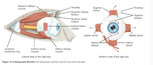
See Figure 21 and Figure 22.

Figure 21: Extraocular movement via cranial nerves. (Illustrator Arina Bogdan)

Figure 22: Extraocular muscles.
(J. Gordon Betts, Kelly A. Young, James A. Wise, Eddie Johnson, Brandon Poe, Dean H. Kruse, Oksana Korol, Jody E. Johnson, Mark Womble, Peter DeSaix. (2022). The Muscular System. In anatomy and physiology 2e. OpenStax. CC BY 4.0 license).
Extraocular eye movement can be assessed using the diagnostic positions test. Use the following steps:
- Sit or stand about 3–4 feet (about two arms-lengths) away from the client, at the same level.
- Ask the client to focus on your nose.
- Normally, the client’s gaze is midline.
- Abnormal findings include deviation of one of the eyes or both: deviation may involve the eye moving inward, outward, up, or down.
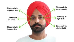
- Place your index finger straight out in front of you and ask the client to focus on your finger. Instruct the client to keep their head still as they follow your finger with their eyes through the six cardinal positions of gaze; see Figure 23). Positions include: diagonally in superior field, laterally at eye level, and diagonally in the inferior field. Move your finger into each of these positions by fully extending your arm: move back to centre each time you move to one of these positions and complete these movements on both sides. Hold your finger in each of these six positions for about two seconds. If the client moves their head, remind them to keep their head still and only follow your finger with their eyes. Ask the client if they experience any double vision. See Video 8.
- Normally, eye movement is smooth, , with no , and no double vision.
- Abnormal findings might include , nystagmus, and double vision. Report any abnormal findings to the physician or nurse practitioner for further evaluation.
- Note the findings:
- Normal findings might be documented as: “Smooth, conjugate movement with no double vision.”
- Abnormal findings might be documented as: “Disconjugate eye movement, nystagmus of left eye with extreme lateral gaze, and double vision noted by client.”

Figure 23: Six diagnostic positions for testing.
Video 8: Extraocular eye movement test. [0.45 seconds].
refers to eyes moving together (in unison).
is repetitive, involuntary eye movement in which the eye looks like it is quivering and can move up-down, side-to-side or in a circular motion.
refers to when eyes do not move together (in unison), but rather one eye moves in a different direction than the other eye.

