Chapter 16 – Vestibulocochlear System Assessment – The Ears
Inspection and Palpation of External Ear
Inspection and palpation of the external ear can be done with the client in any position, but is typically completed with the client sitting in an upright position.
Steps of inspection include (see Video 1):
- Inspect symmetry, lesions, and deformities on or around the ears.
- Normally, the ears are symmetrical with no lesions or deformities. However, one common and normal ear lesion is Darwin’s tubercle (a thickening of or nodule on the middle to upper portion of the helix) (Figure 3).
- In some congenital abnormalities, one ear is significantly smaller or larger, or parts of an ear or the whole ear is not present. Another common deformity is cauliflower ear (Figure 4) caused by a physical trauma to the ear such as a sports injury: disruption to the blood supply causes inflammation and cartilage overgrowth, resulting in a deformity that has the textured appearance of a cauliflower. Ear keloids (Figure 5 and 6) are also a common deformity: these are nodules that can range in size and are firm and rubbery, often caused by minor trauma and scar tissue formation such as with ear-piercing. Ear keloids usually have the same colour as the client’s skin, but can also appear as shades of pink, red, and purple.
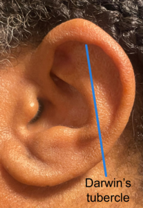
Figure 3: Darwin’s tubercle.
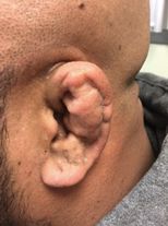
Figure 4: Cauliflower ear. (From: https://jetem.org/cauliflower_ear/cauliflower-ear-photograph-1-jetem-2018/ Cauliflower Ear. Photograph 1. JETem 2018 6. This work is licensed under a Creative Commons Attribution 4.0 International License).
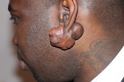
Figure 5: Ear keloid. (By Htirgan, This file is licensed under the Creative Commons Attribution-Share Alike 3.0 Unported license, https://commons.wikimedia.org/wiki/File:Earlobe_Keloid,_Bulky.JPG)
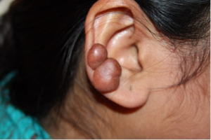
Figure 6: Ear keloid (right image). By Htirgan – Own work, Creative Commons Attribution-Share Alike 3.0 Unported license, https://commons.wikimedia.org/w/index.php?curid=32782666
- Inspect for skin integrity, colour, and swelling on and around the ears.
- Normally, the skin is intact with no signs of discolouration or swelling. If piercings are present, assess for and describe any signs of infection such as erythema and swelling.
- Abnormal findings may include skin that is not intact such as an open ulcer (Figure 7) or the presence of scars. Discolouration of the ear from the client’s baseline is important to evaluate such as erythema (Figure 8) and swollen or darker than the client’s baseline skin colour including shades of purple and blue and turning very black. If present, describe the location and appearance.
- Inspect for bugs and insect bites. Ensure you inspect the crevices of the external ear and behind the ears.
- Normally, no bugs or bites are present.
- If bugs or bites are present, note the location and appearance. For example, lice are bugs that are commonly found among school-age children and are often observed behind the ears.
- Inspect the external auditory meatus and canal for discharge, swelling, discolouration (erythema), and foreign objects.
- Normally, no discharge, swelling, discolouration, or foreign object are present.
- If you observe discharge, swelling, or discolouration, note the appearance (colour), quantity, and location, including which ear. If a foreign object is present, describe the appearance and location, including which ear.
- Inspect for the presence of earwax.
- Normally, some or no earwax is present.
- If earwax is present, note the colour, texture, and quantity.
- Note the findings:
- Normal findings might be documented as: “Ears symmetrical and intact with no lesions, deformities, discolouration, swelling, bugs, discharge, foreign objects, or earwax.”
- Abnormal findings might be documented as: “Left auditory meatus is red and swollen with a yellow sticky discharge” or “Scaly skin rash posterior to right ear” (see example of postauricular eczema). See Figures 9, 10, and 11 for images of an ear infection with discharge.
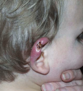
Figure 7: Ulcer. (By Paul D.R. Johnson, Joseph Azuolas, Caroline J. Lavender, Elwyn Wishart, Timothy P. Stinear, John A. Hayman, Lynne Brown, Grant A. Jenkin, and Janet A.M. Fyfe. – Mycobacterium ulcerans in Mosquitoes Captured during Outbreak of Buruli Ulcer, Southeastern Australia. Emerging Infectious Diseases, Volume 13, Number 11.,
This image is a work of the Centers for Disease Control and Prevention, part of the United States Department of Health and Human Services, taken or made as part of an employee’s official duties. As a work of the U.S. federal government, the image is in the public domain,
https://commons.wikimedia.org/w/index.php?curid=34972908)
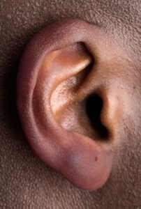
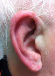
Figure 8: Erythema. (Left image by Tayiba Rahman, CC BY NC. Right image by Giorgio Lambru, Sarah Miller and Manjit S Matharu – “The red ear syndrome”, The Journal of Headache and Pain 2013, 14:83. doi: 10.1186/1129-2377-14-83, CC BY 2.0, https://commons.wikimedia.org/w/index.php?curid=40946332)
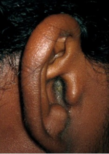
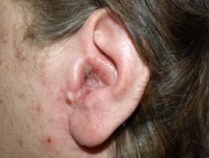
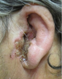
Figures 9, 10, and 11: Ear infection (external otitis [swimmer’s ear]).
Figure 9: (By Grook Da Oger – Own work, Creative Commons Attribution-Share Alike 3.0 Unported license, https://commons.wikimedia.org/w/index.php?curid=16079610)
Figure 10: External otitis. (By Klaus D. Peter, Wiehl, Germany – Own work, Creative Commons Attribution 3.0 Germany license, https://commons.wikimedia.org/w/index.php?curid=9655043)
Figure 11: External otitis – severe case (By James Heilman, MD – Own work, Creative Commons Attribution 3.0 Unported license, https://commons.wikimedia.org/w/index.php?curid=10313999)
Video 1: Ear inspection. [0.59 seconds].
Steps for palpation include (see Video 2):
- Assess one ear at a time. Ask the client if they have any pain as you gently pull the helix upwards and the lobe downwards, and then push on the tragus and the mastoid process.
- Normal findings are no pain.
- Pain is an abnormal finding. If pain is present, assess with the PQRSTU mnemonic.
- Gently palpate and describe any lumps or deformities that you observed.
- Normally, no lumps or deformities are present.
- If a lump or deformity is present, ask the client if it is painful and measure its size, and note any signs of infection such as erythema and swelling. Describe the following characteristics: location on ear(s), colour, texture (smooth, rough, dotted), consistency (soft, firm), and mobility (moveable, fixed).
- Note the findings:
- Normal findings might be documented as: “Client reports no pain when palpating the ear, and no lumps or deformities.”
- Abnormal findings might be documented as: “Firm, rubbery lump at back of ear lobe, 2×2 centimetres. Colour consistent with client’s face.”
Video 2: Ear palpation. [0.26 seconds].
Contextualizing Inclusivity
If a client has ear jewelry, assess for any signs of infection, which can result from non-sterile piercing approaches. Remember to critically reflect on your biases related to ear jewelry: you are likely to observe various types and quantities of ear jewelry on many parts of the ear. Earlobes are a common location, but you may observe jewelry on other areas such as the cartilage of the upper ear and also on the tragus. Some clients may have stretched their ear-piercing holes to accommodate spacers (large round jewelry), Figures 12, 13, and 14. Ear-piercings may be related to culture and spirituality; they are common among women, but may be observed among people of all genders and ages. For example, people of South Asian and Southeast Asian heritage often have their ears pierced before the age of one (Anwar, 2024). Reflect on your own biases and ensure a non-judgemental approach.

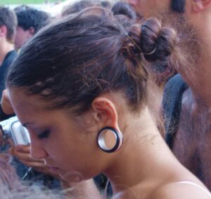
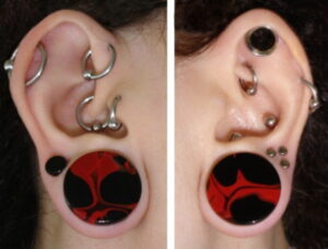
Figure 12: Photo by Sneha Cecil on Unsplash
Figure 13: By Gabriele from Bologna, ITALY – lobe stretching xxl, CC BY 2.0, https://commons.wikimedia.org/w/index.php?curid=5246354
Figure 14: By Lish Daelnar – Lish Daelnar, http://compunction.org, CC BY 3.0 us, https://commons.wikimedia.org/w/index.php?curid=4733704
In young children, it is important to assess the presence and size of ears. Microtia is a congenital deformity of an incompletely formed external ear in which the ear is smaller and sometimes has missing or deformed key features, or the outer ear may be missing altogether (see Figure 15). Macrotia is another congenital deformity in which the external ear is larger than normal and not in proportion to the head; it may be longer and/or wider than normal.
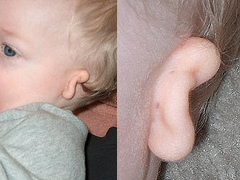
Figure 15: Microtia. (By Mulgamutt at English Wikipedia – Transferred from en.wikipedia to Commons by Filip em using CommonsHelper., Public Domain, https://commons.wikimedia.org/w/index.php?curid=4120960)
Priorities of Care
Report all abnormal findings to the physician or nurse practitioner.
- Erythema, swelling, and discharge are signs of infection and should be reported promptly: an infection needs treatment so it doesn’t lead to permanent damage such as hearing impairment. If the client shows signs of ear infection and/or pain, assess the head and neck lymph nodes: inspection and palpation process.
- Any foreign objects should also be reported promptly. For example, a child might put a small disc battery (button battery) in their ear. This requires immediate action and should be reported immediately, as moisture in the ear can quickly cause batteries to release chemicals, leading to chemical burns, tissue necrosis, tympanic perforation, and/or hearing impairment.
- If a client presents with hypothermia due to extreme cold exposure, assess for findings related to frostbite, which can present as a range of red, purple, pale, blue, and black shades. Assess for pain and paresthesia and numbness or lack of sensation in particular. Slowly rewarm the ears to protect the skin.
- Findings such as lesions should be reported promptly, as they may suggest potential carcinoma. Some clients may tell you they have noticed an open sore that has developed with no physical trauma and has not healed. Any sore that doesn’t heal should be of concern: the healing process depends on the type and severity of the sore, but a typical timeframe to heal is 4 to 6 weeks. Lesions are not a critical finding that require immediate action, but they can be life-threatening and require prompt intervention because they may be curable if diagnosed and treated early.
Activity: Check Your Understanding
References
Anwar, A. (2024). The cultural significance and meaning of ear piercing tradition. https://heyrowan.com/blogs/hey-rowan/how-ear-body-piercing-relates-to-culture-and-tradition

