Chapter 9 – Musculoskeletal System Assessment
Introduction to the Musculoskeletal System
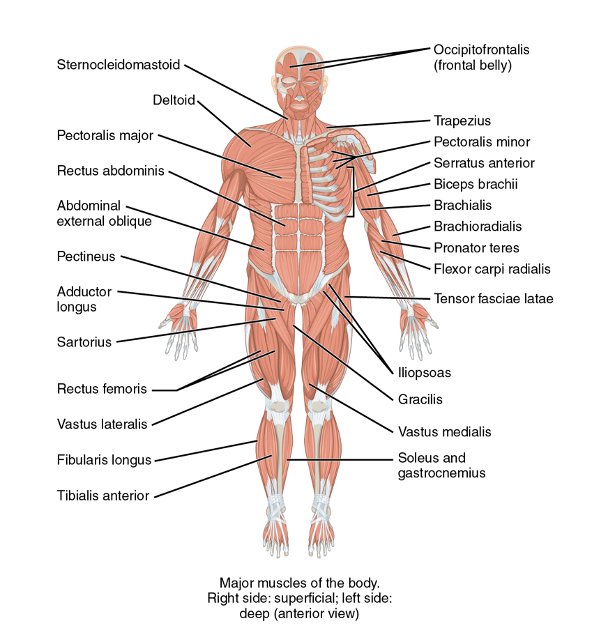
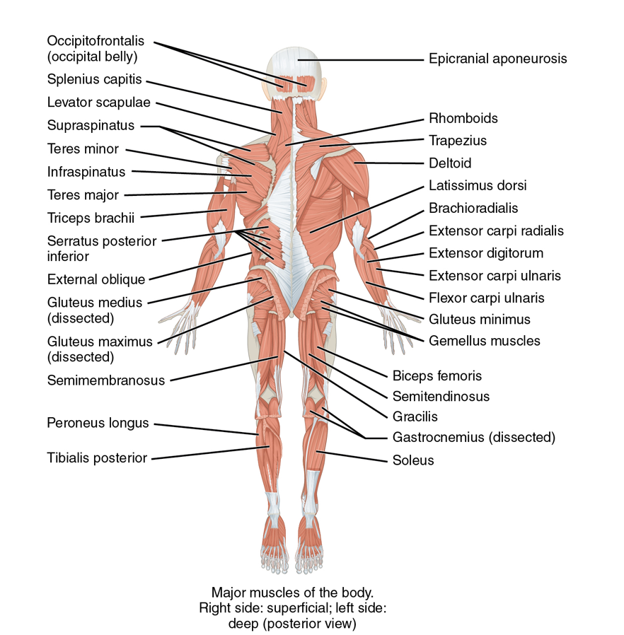
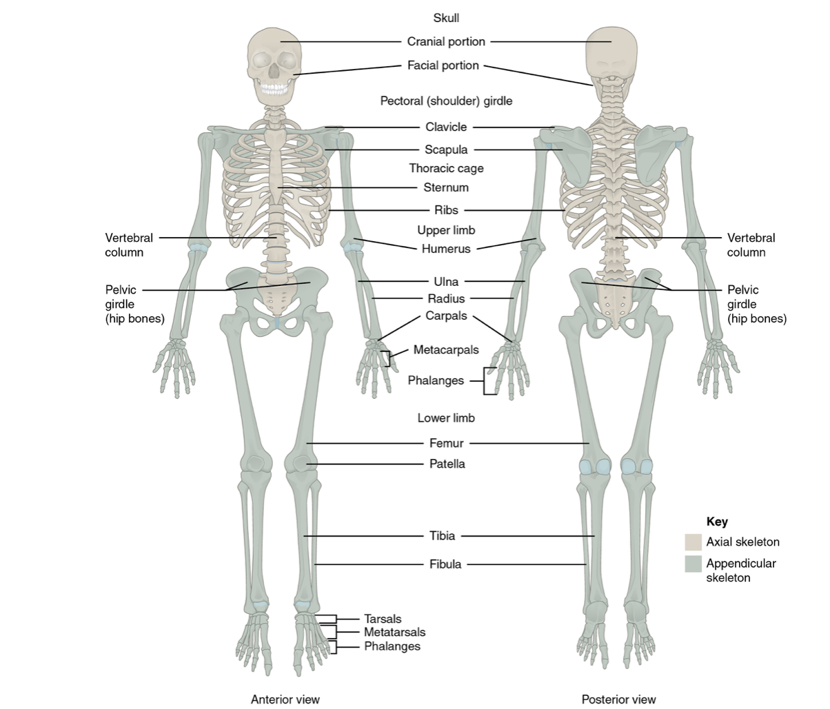
The musculoskeletal system (MSK) is important to assess as it is considered the body’s framework and supportive structure. It has additional roles such as , and mineral and fat storage. See Figures 1 and 2 for an anatomical overview of the MSK system.
Your assessment provides information about the functioning of this system and potential cues that require your action.
Musculoskeletal System Components
The main components of the MSK system include the skeleton:
- Skull, including the cranial and facial bones.
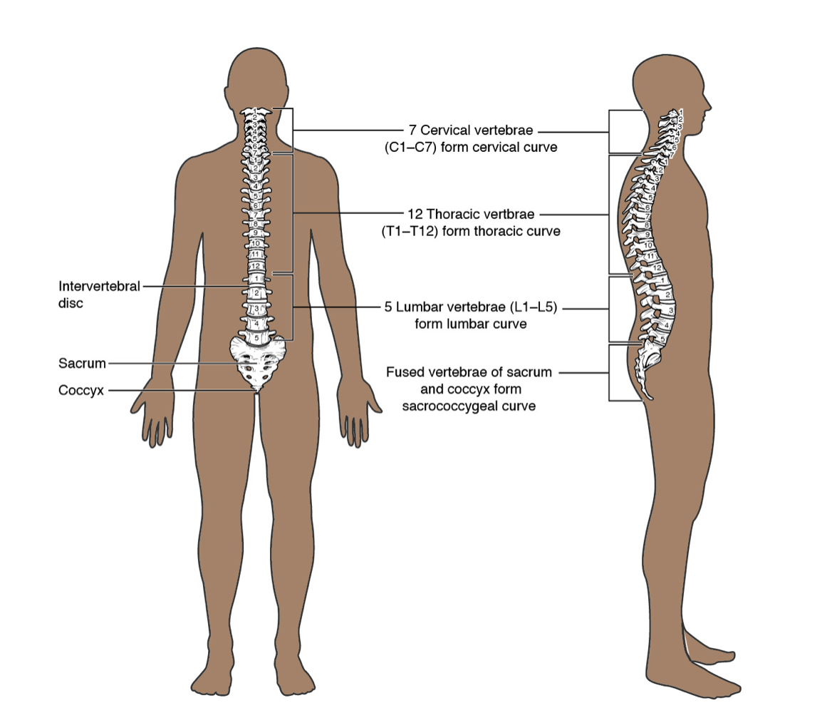
- Vertebral column with intervertebral discs.
- Thoracic cage with sternum and ribs.
- Clavicle and scapula.
- Upper and lower limb bones.
- Hip/pelvis bones.
The skeleton also includes cartilage in particular areas of the body such as the nose, ears, , and in joints (e.g., shoulder, hips, knees, elbows, wrists/hands, and ankles/feet). See Figure 3 for an anatomical overview of the articular cartilage and synovial joint.
The skeleton is supported by muscles. Skeletal muscles specifically attach to the bones of the skeleton and provide support. The MSK system is connected and supported by joints. These joints include articular cartilage, ligaments, and tendons to connect the MSK system together. Many joints have a cavity with synovial fluid between the articulating joints that permits mobility, including the shoulders, elbows, wrists/fingers, hips, knees, and ankles/toes. Other joints do not have a cavity with synovial fluid including cranial sutures (which are not moveable) and the vertebral column (which is slightly moveable). Bones are connected by ligaments, and tendons connect muscle to bone.


Figure 1: Muscular system (anterior view on top image, posterior view on bottom image)
Attribution: Building a Medical Terminology Foundation by Kimberlee Carter and Marie Rutherford is licensed under a Creative Commons Attribution 4.0 International License, except where otherwise noted.


Figure 2: Skeletal system
Attribution: Building a Medical Terminology Foundation by Kimberlee Carter and Marie Rutherford is licensed under a Creative Commons Attribution 4.0 International License, except where otherwise noted.
 Figure 3: Articular cartilage and synovial joint.
Attribution: Building a Medical Terminology Foundation by Kimberlee Carter and Marie Rutherford is licensed under a Creative Commons Attribution 4.0 International License, except where otherwise noted.
You have already learned about the anatomy and physiology of the MSK system, but check out these helpful videos on the muscular system explained in six minutes and the skeletal system explained in seven minutes.
Clinical Tip
Your MSK assessment will begin when you first meet the client. Remember to incorporate the concept of functional health by assessing the client’s physical capacity to participate in daily activities. For example, a functional assessment includes assessing day-to-day activities such as walking into the room, moving from standing to sitting, signing a consent form, or changing into a hospital gown. Always compare bilaterally during the assessment. Ask clients if they have noticed any changes in their movement and if they have any questions or concerns. Your findings will help to guide your assessment throughout the examination, as well as provide opportunities to incorporate health promotion and prevention education.
Knowledge Bites
The MSK system is influenced by the neurological system. For example, muscle functioning (e.g., movement and strength) is innervated by the cranial nerves (12 pairs of nerves that send electrical signals from the brain to the body) and the spinal nerves.
Activity: Check Your Understanding
refers to blood cell formation.
connects the sternum to the ribs and helps with motion to expand the chest during respiration.
is flexible connective tissue covering the end of bones in synovial joints and help the bones glide over one another.

