
Chapter 4 – Cardiovascular System Assessment
Inspection of the Precordium
The precordium is the region on the chest wall that overlays the heart area. The base of the heart is located in the region of the second intercostal space and the apex is located in the region of the fifth intercostal space on the left side. As you inspect the chest wall, you will collect data that provides information about cardiac function.
Inspection of Precordium
1. Place the client in supine position with their head on a pillow.
2. Provide draping.
3. Use tangential lighting with a penlight across the heart area.
4. Inspect the base of the heart, left sternal border, and apex of heart. You may do this in sequence so that you are only exposing the area that you are assessing and then move on to the next area. See Figure 4.6 and Video 4.6.
5. Observe for the presence or absence of against the chest wall and note the location.
- Pulsations: These are gentle flickers observed on the skin of the chest wall. They can be considered normal when observed on clients with thin chest walls. Otherwise, these are cues that require further assessment because they may be suggestive of increased cardiac workload associated with conditions such as an enlarged heart.
- Heaves: These are a more forceful movement (a thrust) observed on the skin over the chest wall. These cues require further assessment because they also may be suggestive of increased cardiac workload such as an enlarged heart.
6. Note the finding
- Normal findings might be documented as: “No cardiac impulses observed against chest wall.”
- Abnormal findings might be documented as (example): “Gentle pulsation observed at apex.” or “Heave observed at apex.”
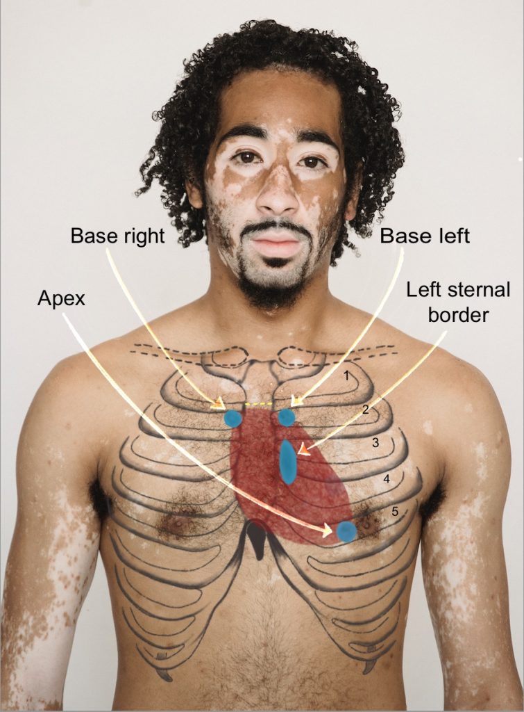
Figure 4.6: Location of the base of the heart, left sternal border, and apex of heart
Photo by Armin Rimoldi from Pexels (image was cropped and illustrated upon for the purposes of this chapter)
Video 4.6: Inspection of precordium
Knowledge Bites – Pathophysiology
Impulses observed on the chest wall should always be assessed further because they are suggestive of some sort of increased workload of the heart and/or vasculature (such as the aorta). A heave can be associated with cardiac hypertrophy (enlargement of the heart muscle) or a dissecting aorta (a tear in the inner arterial wall that can lead to a rupturing of the aorta). These can be caused by many conditions such as hypertension, valvular disease, septal defects, and infarction. Your assessment can contribute to the database about whether the impulse is benign, such as may appear with a client who has a thin chest wall, or whether the impulse may have a pathophysiological cause.
Activity: Check Your Understanding
are movements visualized on the chest wall.
