
Chapter 3 – Respiratory System Assessment
Posterior and Lateral Thorax – Inspection
1. Inspect for symmetry of thorax and chest expansion.
- Compare the left and right sides of the thorax. Are the shoulders, scapula, and ribs on symmetrical upon observation?
- When the client breathes, does the left and right sides of the thorax expand and recoil symmetrically? Also, test for symmetrical chest expansion by placing your thumbs (on either side of vertebra) just below the inferior border of the scapula (about T9/T10) with fingers stretched out to the lateral sides of the thorax. Pinch a piece of skin in between your thumbs and ask the client to take a big breath in and out. If the thumbs move equally apart and back together at the same time upon inspiration and expiration, this indicates symmetrical expansion. Make a note if the expansion is asymmetrical (one side has limited expansion, lags in expansion, or does not expand). This can happen when one lung cannot expand due to conditions that involve inflammation and air between the lungs and chest wall (i.e., pneumothorax) or partial or complete collapse of the alveoli (i.e., atelectasis). You may also notice that in pregnant women, the thorax shortens while the costal angle widens to accommodate the enlarging uterus: this is normal.
2. Inspect the spinous process, ribs, , masses, and swelling.
- Do the spinous processes appear in a straight line down the vertebral line of the posterior thorax?
- Are the ribs sloping downwards?
- Do you observe any deformities, masses or swelling?
3. Inspect skin colour and skin integrity
- Is the skin colour consistent across the posterior and lateral thorax?
- Do you notice any skin discolouration?
- Do you notice any scars? If so, ask the client the cause. For example, a client may have a scar from lung surgery such as a , and this surgery can lead to no air entry when you auscultate the lungs.
4. Inspect the anteroposterior to transverse diameter of the thorax. See Figure 3.10. The transverse diameter is shown in the first image across the chest. The anteroposterior diameter is show in the second image from front to back. In most adults, the anteroposterior to transverse diameter ratio is about 1:2.
- The ratio will be closer to equal (1:1) when a client has conditions that give rise to hyperinflated lungs (e.g., emphysema). Also, a 1:1 ratio is usually present in children younger than two years of age.

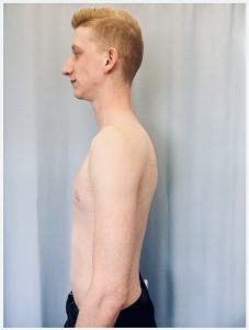
Figure 3.10: Anteroposterior to transverse diameter
5. Note the findings
- Normal findings might be documented as: “Symmetrical posterior thorax with symmetrical chest expansion, no thorax deformities or masses, spinous process in a straight line, no skin discolouration, anteroposterior to transverse diameter 1:2.”
- Abnormal findings might be documented as: “Horizontal ribs with a 1:1 anteroposterior to transverse diameter.”
Priorities of Care
None of the abnormal findings on their own are considered critical findings. However, if you notice asymmetrical lung expansion, be aware that this is associated with decreased ventilation. You should investigate whether there is decreased or absent air entry in the affected lung, which may be lagging behind or not expanding at all. Examples of underlying pathologies can include , , , or partial or complete obstruction of the airway on the side.
Activity: Check Your Understanding
are abnormal malformations of the body.
is the surgical removal of one of the lobes of the lung.
is a collapsed lung as a result of air entering the pleural space between the visceral and pleural layers.
is a partial collapse of lung when the alveoli become deflated usually caused by shallow breathing following surgery, blocked airways, or insufficient surfactant.
refers to excessive fluid accumulation in the pleural space between the visceral and parietal pleura.
refers to same side.
