
Peripheral Vascular System Assessment
Introduction to the Peripheral Vascular System
The peripheral vascular system (PVS) is a continuous network of blood vessels that carry oxygenated blood away from the heart to the periphery and carry deoxygenated blood back to the heart and to the lungs for reoxygenation. This system is important in the perfusion and oxygenation of tissues in the periphery. If this system is not functioning properly, perfusion issues can arise including and tissue damage.
Assessment of the PVS provides information about the functioning of this system and cues that may require action.
Peripheral Vascular System Components
The main components of the PVS (see Table 1 and Figures 1 and 2) include:
- Arteries: vessels that carry blood away from the heart to the periphery. Blood in the arteries is referred to as arterial blood. The multiple layers of the arteries are strong and elastic and can dilate (expand in circumference) and recoil (decrease or return to normal size) in relation with cardiac and . These high-pressure vessels dilate when the heart contracts and pumps blood out into them and then recoil to push blood through the arteries, creating a wave of blood through the vessels (felt as a pulse).
- Veins: vessels that carry blood back to the heart from the periphery. Blood in the veins is referred to as venous blood. The walls of these vessels are thin in comparison to arteries but have good stretching capacity, so they can acclimate to larger volumes of fluid (blood) when needed. Veins are low-pressure vessels because they are further away from the heart than arteries. Cardiac systole (contraction of the heart) does not assist with the forward movement of venous blood the way it does with arterial blood. Rather, forward movement of venous blood is mainly achieved through contraction of the skeletal muscles surrounding these veins and in the veins that maintain unidirectional blood flow and prevent backward flow of blood.
- Capillaries: small blood vessels that connect the arteries to the veins. Their main function is to facilitate the exchange of materials between blood and body tissues (e.g., muscles, kidneys, liver). They deliver blood and its components (nutrients and oxygen) to tissues throughout the body and transport waste products.
You have already learned about the anatomy and physiology of the PVS assessment: see this video for a quick overview: https://youtu.be/v43ej5lCeBo
Table 1: Comparison of arteries and veins. (Image from Anatomy and Physiology [on OpenStax] by Betts et al., used under a CC BY 4.0 international license. Download and access this book for free at https://openstax.org/books/anatomy-and-physiology/pages/1-introduction)
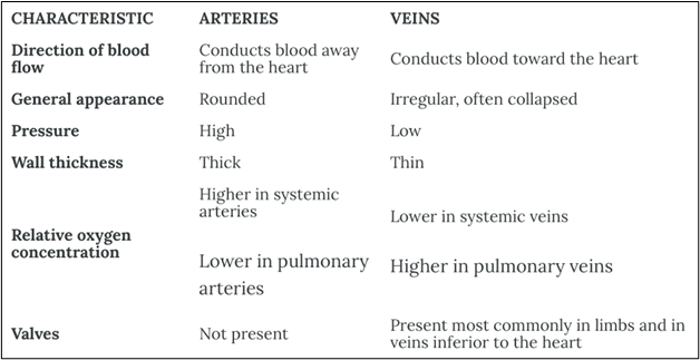
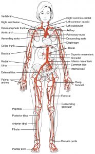
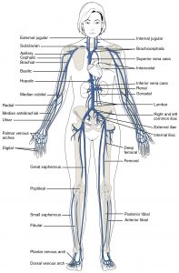
Figure 1: Peripheral vascular system anatomy. (Image from Anatomy and Physiology [on OpenStax] by Betts et al., used under a CC BY 4.0 international license. Download and access this book for free at https://openstax.org/books/anatomy-and-physiology/pages/1-introduction)
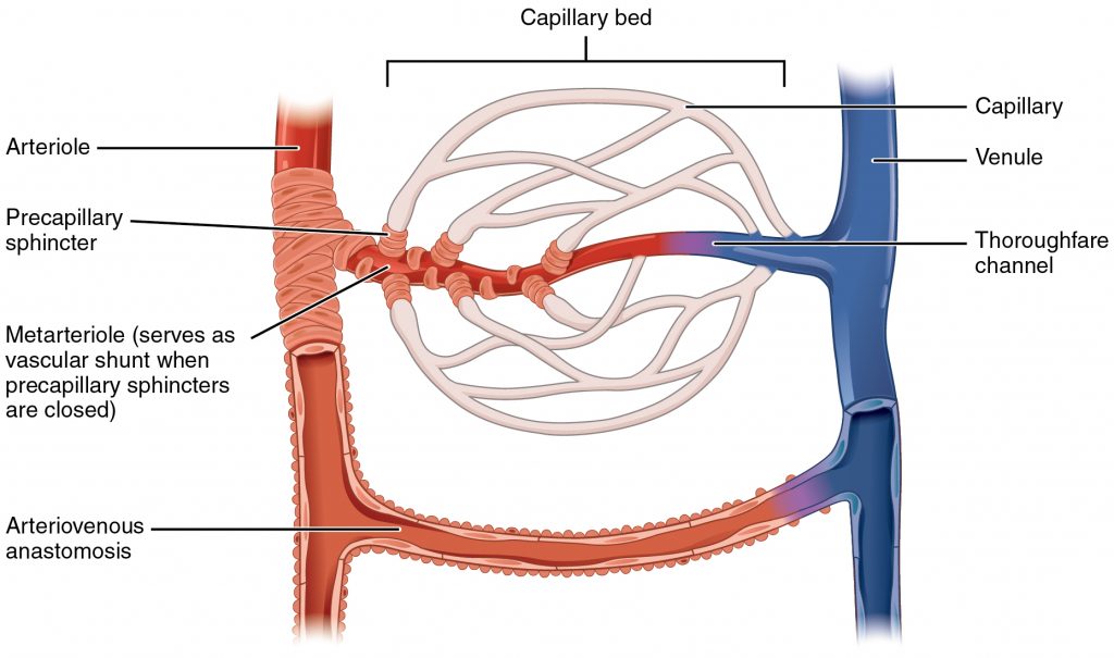
Figure 2: Capillaries. (Image from Anatomy and Physiology [on OpenStax] by Betts et al., used under a CC BY 4.0 international license. Download and access this book for free at https://openstax.org/books/anatomy-and-physiology/pages/1-introduction)
Clinical Tip
The PVS is interconnected with many other body systems, so it is rarely assessed in isolation. When attempting to make sense of cues, nurses commonly assess other body systems including cardiovascular, integumentary, lymphatic, and musculoskeletal. See Figure 3 for related systems. For example, the PVS might be assessed along with a cardiovascular assessment to assess perfusion and also with the musculoskeletal system to assess potential blood flow interruptions. The PVS is also closely related to the lymphatic system and connected via the capillaries. In addition, many PVS conditions affect the skin, thus the integumentary system is often assessed.
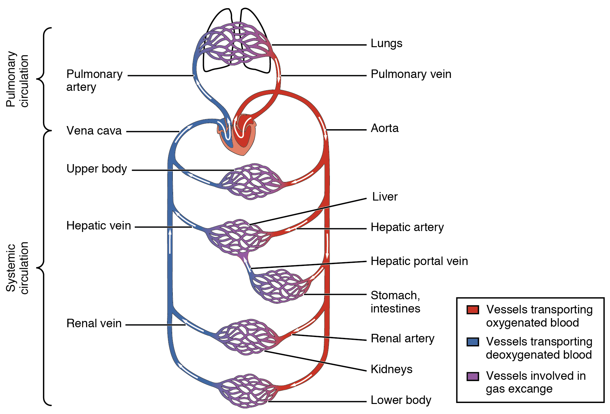
Figure 3: Pulmonary and systemic circulation.
(Image from Anatomy and Physiology [on OpenStax] by Betts et al., used under a CC BY 4.0 international license. Download and access this book for free at https://openstax.org/books/anatomy-and-physiology/pages/1-introduction)
Activity: Check your Understanding
refers to low levels of oxygen in tissues and organs.
is the cardiac phase when the heart is full of blood and the ventricles contract ejecting blood into the arteries.
is the cardiac phase when the heart's chambers are at their lowest pressure and filling with blood.
are valves within the veins.
